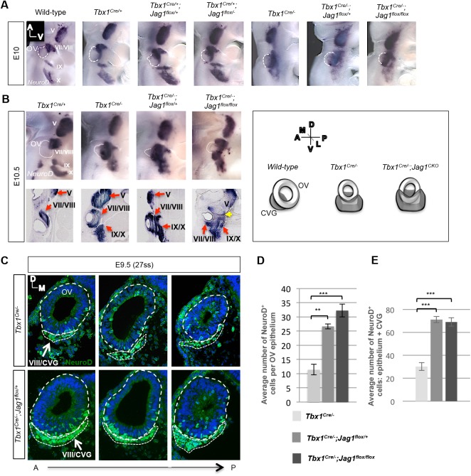
Fig. 4.
Inactivation of Tbx1 and Jag1 with Tbx1Cre results in expanded proneural gene expression. (A,B) Whole-mount in situ hybridization was performed on genotypes shown, using an antisense probe for NeuroD at E10 (A) and E10.5 (B). There is a synergistic increase in expression in the CVG (cranial ganglion VIII) as Tbx1 and Jag1 dosage decreases. There is a similar increase in expression in the other cranial ganglia as well (VII, IX, X), shown in both whole mounts and sections. Fusion of all the ganglia with the Vth ganglion occurs in Tbx1Cre/−;Jag1flox/flox mutants, indicated by the yellow arrow. Adjacent is a schematic depicting the phenotype observed in Tbx1 single and Tbx1;Jag1 double mutants. (C) Immunofluorescence on transverse sections with a NeuroD antibody. Both the CVG and OV epithelium are outlined with a white dashed line. The mean total numbers of NeuroD+ cells in the OV epithelium alone (D) and the OV epithelium in combination with the CVG (E) were significantly greater in double mutant OVs as compared to Tbx1Cre/− OVs. Note: three embryos and six OVs per genotype were used for each group. Embryos were stage-matched at 27ss. Data are mean±s.e.m. **P<0.005; ***P<0.001.

