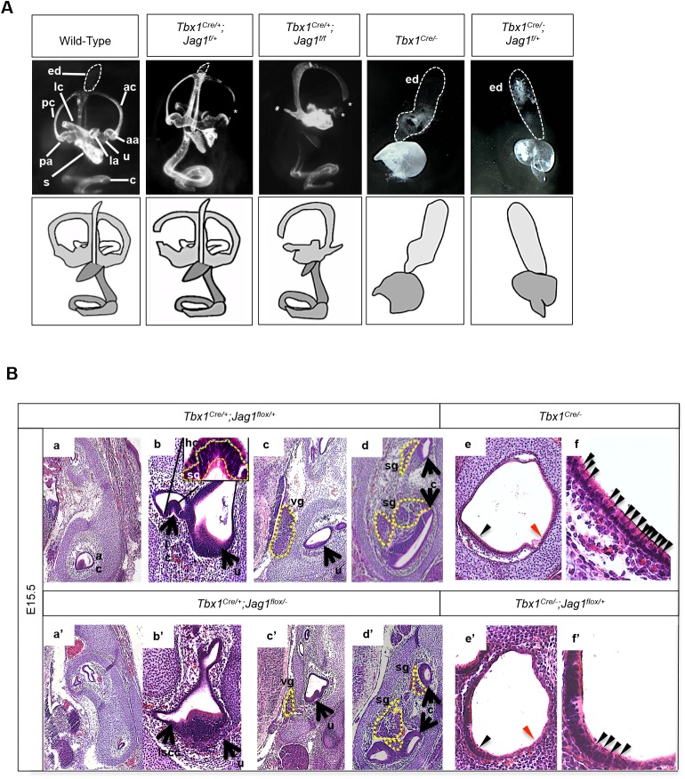
Fig. 6.
Gross morphological inner ear defects in Tbx1;Jag1 compound mutant embryos at E15.5. (A) Paintfilling of E15.5, Tbx1Cre/+;Jag1flox/+, Tbx1Cre/+;Jag1flox/flox, Tbx1Cre/−, Tbx1Cre/−;Jag1flox/+ and wild-type control inner ears. Tbx1Cre/+;Jagflox/+ ears are relatively normal but present with narrowing of the canals, particularly the anterior canal (white asterisk). Tbx1Cre/+;Jagflox/flox ears display incomplete development of the anterior semicircular canal (ac), posterior semicircular canal (pc) and lateral semicircular canal (lc), as well as their associated ampullae, respectively (aa, pa, la) (white asterisks). Tbx1Cre/− mutant ears have an enlarged endolymphatic duct (ed) (white tracing), while the rest of the inner ear does not fully develop and remains a vesicle with a posterior protrusion. Tbx1Cre/−;Jagflox/+ mutant ears are similar to Tbx1Cre/− ears but the vesicle is smaller and the protrusion is extended ventrally. (B) Transverse histological sections stained by H&E. All three cristae develop normally in Tbx1Cre/+;Jag1flox/+ embryos (a,b). The anterior crista (ac) and lateral crista (lc) attached to the utricle (u) are shown. An image of b at a higher magnification in the inset shows rows of hair cells (hc) and support cells (sc) that develop normally. All three cristae are missing from Tbx1Cre/+;Jag1flox/flox ears (a′,b′). Vestibular (vg) and spiral ganglia (sg) appear to form normally in Tbx1Cre/+;Jag1flox/+ embryos (c,d). The vg is noticeably smaller in Tbx1Cre/+;Jag1flox/flox embryos while the sg appears to develop normally (c′,d′). The inner ear rudiment (the vesicle ventral of the endolymphatic duct) in Tbx1Cre/− mutants overall are mostly lacking in hair cells, particularly in lateral regions (red arrowhead); however, the medial ventral wall of the vesicle is fairly densely populated with hair cells (black arrowhead) (e). An image of e at a higher magnification is shown in f. Tbx1Cre/−;Jag1flox/+ ears are similar to Tbx1Cre/− ears in that they are mostly lacking hair cells, particularly in lateral regions (red arrowhead) (e′). Medial regions are populated by some hair cells, but much more sparsely. An image of e′ at a higher magnification is shown in f′.

