
Fig. 5.
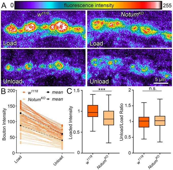
Notum loss alters presynaptic differentiation via vesicle trafficking. (A) Representative synaptic bouton images of FM 1-43 dye imaging with depolarization-induced loading (top) and unloading (bottom) in w1118 control (left) and NotumKO (right). Fluorescent intensity is represented as a heat map. (B) Sample quantification of FM1-43 dye loaded and unloaded bouton fluorescence for all boutons from a single NMJ of each genotype. (C) Box-and-whisker plot quantification of the loaded bouton fluorescence (left) and unload/load ratio (right) for all boutons. ***P≤0.001; n.s. not significant.