Fig. 6.
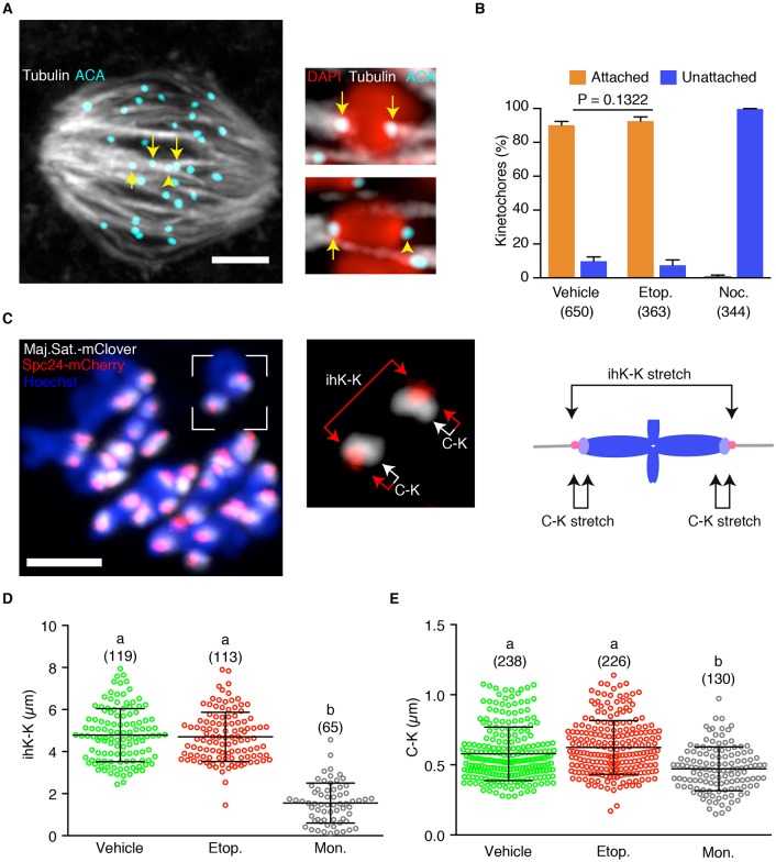
DNA damage does not reduce kinetochore-microtubule attachment or tension. (A) K-fibres immunostained for tubulin and anti-centromere antigen (ACA; counterstained with DAPI). Two bivalents are shown at higher magnification, showing examples of attached (arrows) and unattached (arrowhead) kinetochores. (B) Frequency of kinetochore attachment to k-fibres following addition of etoposide, nocodazole or vehicle (0.1% DMSO). Numbers of kinetochores assessed are shown in parentheses. Statistical test was Fisher's exact, error bars represent 95% confidence intervals. (C) Bivalents in an oocyte expressing Spc24-mCherry, a TALE protein against the major satellite repeat (Maj. Sat.-mClover), and counterstained with Hoechst; at 7 h after NEB. Right-hand image (detail of the boxed area on the left) and diagram show the measurements made on each bivalent: inter-homologue kinetochore stretch (ihK-K) and centromere-kinetochore stretch (C-K). (D,E) Measures of ihK-K (D) and C-K (E) following treatment with vehicle (DMSO, 0.1%), etoposide or monastrol (100 µM). Error bars indicate s.d.; number of measurements are in parentheses, different letters indicate statistically significant differences (P<0.05; ANOVA with Tukey's multiple comparison test). Error bars indicate s.d. Scale bars: 10 μm in A; 5 μm in C.

