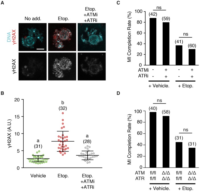
Fig. 7.
Meiotic DNA damage-induced SAC activation is independent of ATM and ATR kinases. (A) Representative γH2AX staining in the nuclei of oocytes before NEB, following addition of etoposide, or etoposide with ATMi (KU55933) and ATRi (ATR kinase inhibitor II). Scale bar: 5 µm. (B) Quantification of γH2AX levels as shown in A. Number of oocytes measured is shown in parentheses. Different letters indicate significant difference (P<0.0001; ANOVA with Dunn's multiple comparison test). (C) Percentage of oocytes completing MI following treatment with either etoposide or DMSO vehicle before NEB. Groups were matured in the presence or absence of ATMi and ATRi, and scored for polar body extrusion. (D) Oocytes from mice that were conditional double knockouts for ATR and ATM, or floxed littermate controls, were exposed to etoposide or a vehicle control, and assessed for completion of MI. (C,D) Number of oocytes used indicated in parentheses, statistical test used was Fisher's exact (ns, not significant). Error bars indicate s.d.

