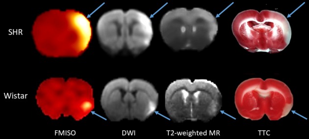
Fig 2. Illustrative coregistered FMISO SUV, DWI and T2-weighted MRI images for the same coronal cut in one spontaneously hypertensive rat and one Wistar rat, and approximately matched TTC coronal slice.

These data illustrate the topographic congruence of the ischemic lesions (arrows) among the four imaging modalities. As TTC sections are not amenable to coregistration with PET and MR images due to differences in slice angle and thickness and potential post-mortem geometrical distortion, approximately same coronal level TTC sections as for the in vivo images are shown for illustrative purposes only.
