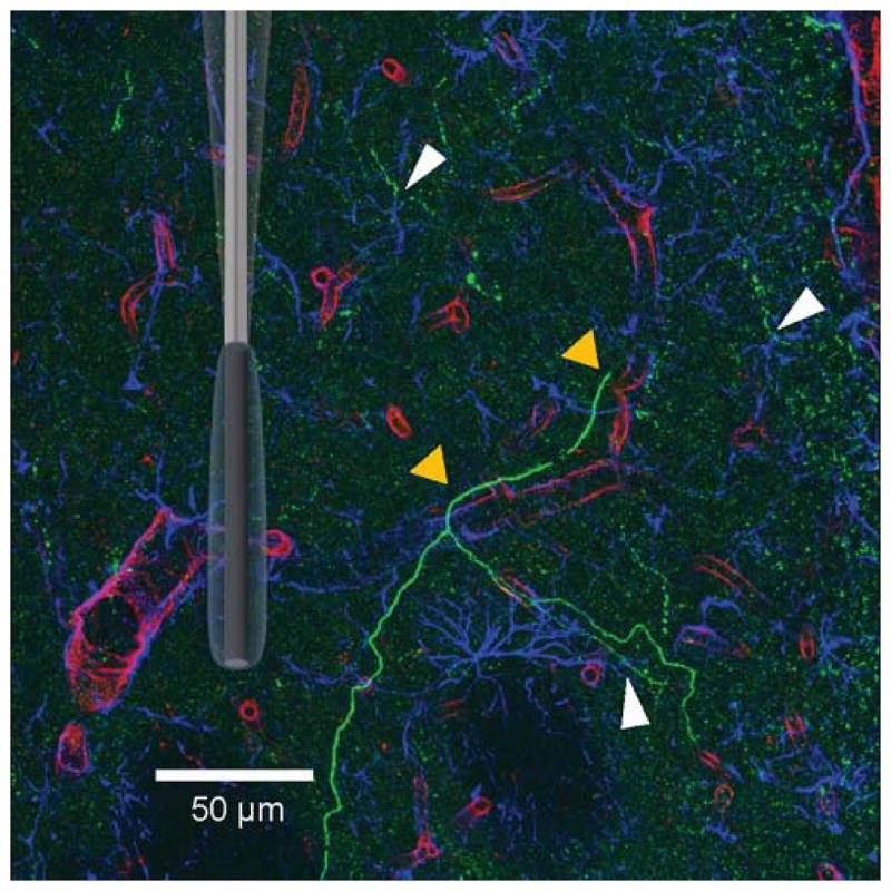
Figure 1.

Triple-Fluorescence Labeling in the Rat Dorsal Striatum. Confocal laser image (275 x 275 μm) taken from a 40 μm-thick tissue slice. Sites where dopaminergic axonal fibers (green; TH label) interact with blood vessels (red; lectin label) and/ or astrocytes (blue; GFAP label) are indicated by the triangles (yellow and white, respectively). A randomly oriented and scaled schematic of a GOx EME is superimposed to demonstrate its relative size in this environment. Scale bar is 50 μm.
