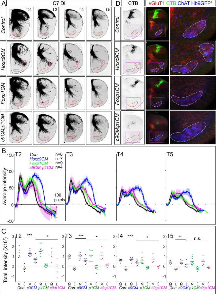
Figure 2. MN Columnar Identity Shapes Sensory-Motor Connectivity.
(A) DiI tracing from DRG C7 analyzed at thoracic segments T2–T5 in indicated mouse mutants at P0. In Hoxc9CM; Foxp1CM double mutants and Foxp1CM mice, SNs do not project to thoracic MNs. (B) Quantification of DiI traced DRG C7 sensory afferents in ventrolateral quadrant of segments T2–T5. Number of animals analyzed: Control, n=6 (P0, n=6); Hoxc9CM, n=7 (P0, n=7); Foxp1CM, n=9 (E18.5, n=3; P0, n=6); Hoxc9CM; Foxp1CM, n=4 (P0, n=4). (C) Quantification of total DiI pixel intensity in medial and lateral regions of the spinal cord. Pixel intensities in lateral position of Hoxc9CM; Foxp1CM and Foxp1CM mice are similar to controls. ***p=0.0004, *p=0.0219, T2; ***p=0.0001, *p=0.0274, T3; ***p=0.0002, *p=0.0192, T4; **p=0.0025, ns=0.0747, T5. (D) Analysis of triceps sensory terminals in segment T3 at P5. In Hoxc9CM; Foxp1CM mice no CTB+ terminals are observed in the ventral spinal cord. n=3 each genotype. See also Figure S2.

