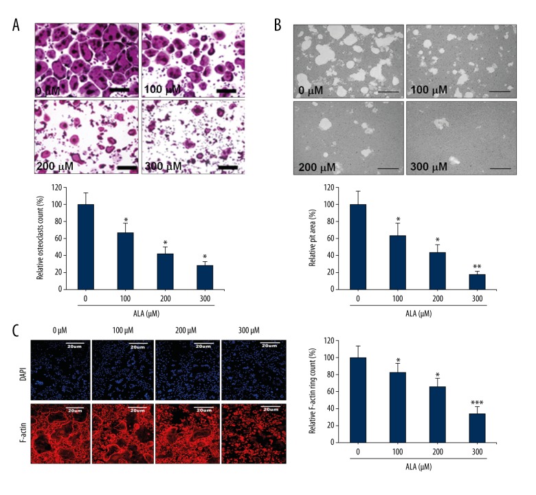
Figure 1.
Effect of ALA on osteoclast differentiation and maturation. (A) BMMs were cocultured with M-CSF (30 ng/mL) and RANKL (100 ng/mL) with indicated concentrations of ALA for 3 days. TRAP-positive multinucleated cells were counted as osteoclasts (bar graph). * P<0.008 vs. control group. (B) Mature osteoclasts seeded on hydroxyapatite-coated plates were treated with indicated concentrations of ALA for 24 h. Cells were imaged and pit areas were visualized and quantified. * P<0.006, ** P=0.002 vs. control. (C) BMMs were cultured and treated with ALA and stained with DAPI with phalloidin. Fluorescence imaging was performed and for visualization and quantification of F-actin rings. * P<0.004, *** P=0.001 vs. control.

