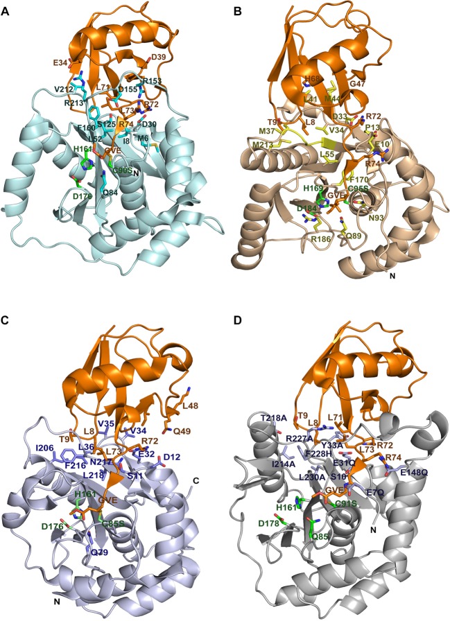
Figure 1. Structure of UCHs bound to the suicide substrate UbVMe.
Ribbon representation of the structure of UCHs–UbVMe complex (A) UCHL1 (3IFW) shown in pale cyan and residues interacting with ubiquitin are highlighted in cyan. (B) UCHL3 (1XD3) shown in wheat with residues interacting with ubiquitin highlighted in yellow (C) TsUCHL5 (4I6N) represented in light blue with residues interacting with ubiquitin highlighted in blue. (D) Structural mapping of predicted ubiquitin-interacting residues of BAP1 onto the UCHL5-based modeled structure of BAP1N in complex with UbVMe. BAP1N is colored gray, with predicted ubiquitin-interacting residues highlighted in black. In all structures UbVMe is shown in orange, catalytic residues are highlighted in green. Covalent bond formed between VMe and catalytic cysteine is shown in yellow. The panel is made using PyMOL (www.pymol.org).

