
Fig. 1.
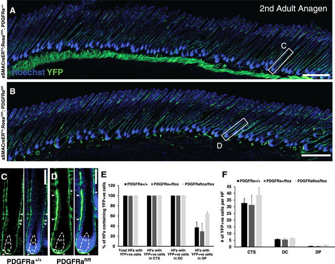
In vivo fate mapping of Pdgfrα-deficient hfDSCs during second adult anagen. a Skin from αSMACreERT2 :RosaYFP :Pdgfrα +/+ or b Pdgfrα flox/flox animals after Cre-induced recombination. The backs of the animals were depilated at P21 (telogen) and tamoxifen was administered by gavage at P21, P23, and P25 (1 mg/day/animal). The subsequent anagen phase was documented and compared between tamoxifen-treated wild-type (n = 3), heterozygous (n = 4), or Pdgfra flox/flox (n = 3), carrying Cre animals. Skin was harvested during second anagen (P33-P35). c Magnified view of an individual HF from Pdgfrα +/+ skin (white box in (a)) showing YFP+ve cells (green, arrowheads) in each of the dermal compartments of the HF, i.e., CTS, DC, and DP outlined by dashed lines. d Magnification of a HF from Pdgfrα flox/flox skin (white box in (b)) for comparison. e Percentage of HFs containing YFP+ve cells. Total number of YFP+ve HFs per genotype are represented, as well as the percentages of HFs with YFP+ve cells present in each dermal compartment. f Mean number of YFP+ve cells per HF present in each component of the dermal compartment in Pdgfrα +/+ ; Pdgfr +/flox and Pdgfrα flox/flox animals. Data are mean ± SEM (n = 10 animals). Kruskal–Wallis test (p = n.s.). Scale bar represents 500 (a, b) or 50 µm (c, d). See also Fig. S1