. 2017 Nov 1;6:e29388. doi: 10.7554/eLife.29388

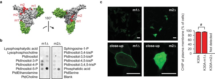
Figure 3. A positively charged patch is required for CHIP binding to phospholipids.

(a) Surface representation of CHIP dimer (PDB entry 2c2l) with TPR domains colored green. m1, m2, positively charged patches colored red. (b) m2-mutation (m2Δ) affects CHIP interaction with phospholipids in vitro as determined by lipid-binding assay. One representative out of three independent experiments is shown. (c) m2Δ CHIP loses association with membranes in vivo. Scale bar 20 μm. #, no statistically significant difference according to chi-square analysis; N = 3 independent experiments (mean ± SD).

Figure 3.

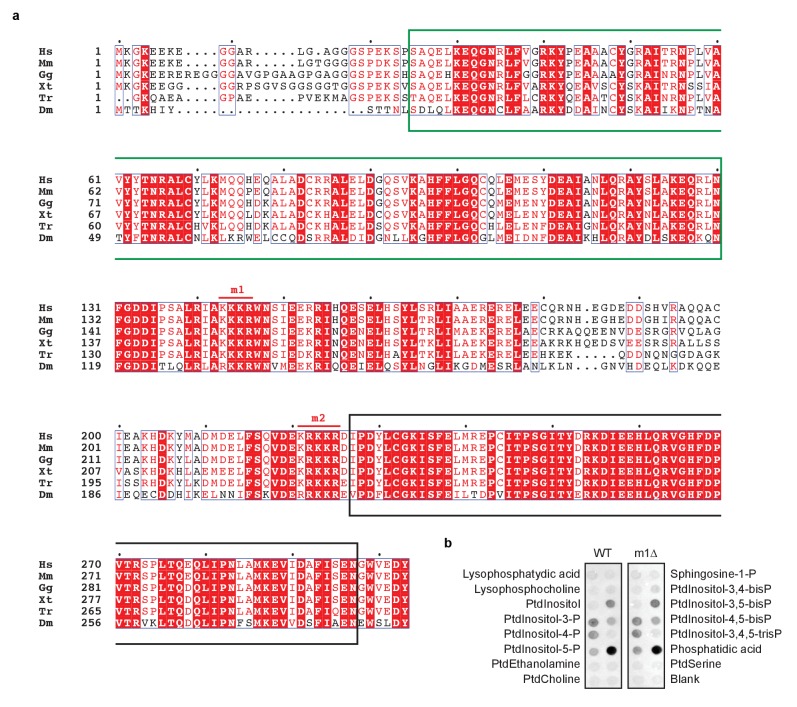
Figure 3—figure supplement 1. Positively charged patches in CHIP are conserved.

Figure 3—figure supplement 1.

(a) Alignment of CHIP sequences from human (Hs, Homo sapiens), mouse (Ms, Mus musculus), chicken (Gg, Gallus gallus), xenopus (Xt, Xenopus tropicalis), fugu (Tr, Takifugu rubripes), and fruitfly (Dm, Drosophila melanogaster). TPR domain and U-box are marked by green and black boxes, respectively. Two positively charged patches are conserved and labelled m1 and m2. (b) Purified wild-type CHIP (WT) and its m1-mutated variant (m1Δ) show similar phospholipid binding specificity as determined by lipid-binding assay. One representative out of three experiments is shown.