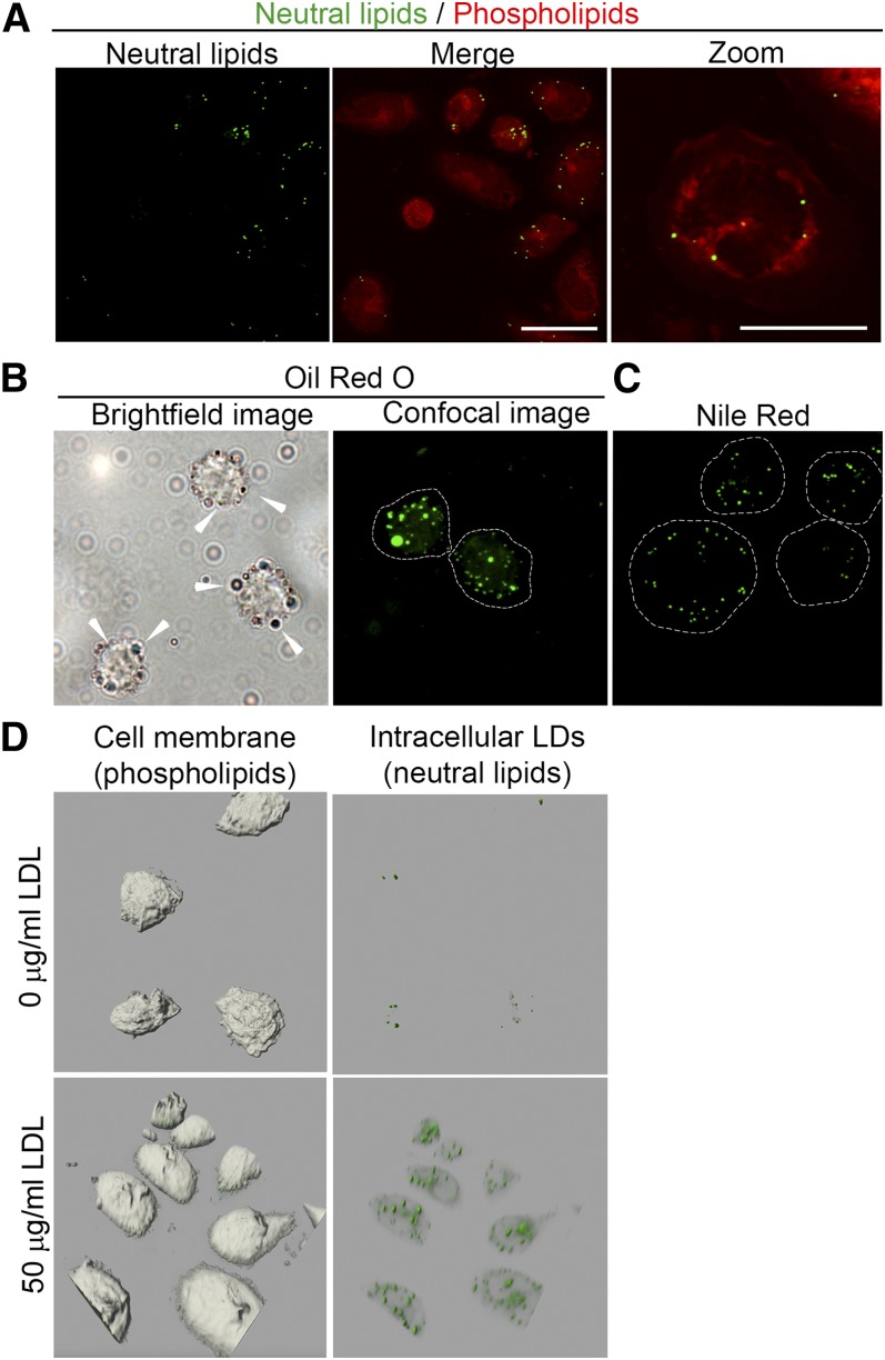
Fig. 1.
LD detection by NR in human peripheral monocytes. A: NR-stained monocytes. Neutral lipids (green spheroids; left image) can be discriminated from phospholipids (red; middle image and right image; scale bars, 20 and 10 μm, respectively). B: Representative images of ORO-stained monocytes incubated with 50 μg/ml LDL for 1 h. The left image shows LDs by bright-field microscopy; LDs are indicated with white arrows. The right image was obtained by confocal microscopy. LDs are shown in green. C: NR-stained monocytes treated with 50 μg/ml LDL for 1 h. LDs are shown in green. Scale bar, 10 μm. D: The 3D images of monocytes (gray bodies shown on left images) untreated (upper images) and treated (lower images) for 1 h with 50 μg/ml LDL. LDs are shown in green.

