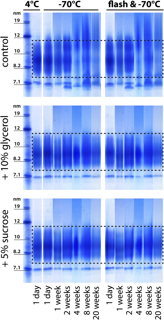
Fig. 5.
Flash-freezing of HDL. HDL was isolated from the serum of four healthy donors by density gradient ultracentrifugation and pooled for electrophoresis analysis. HDL with or without the cryoprotectants (10% glycerol or 5% sucrose) was either frozen directly at −70°C or flash-frozen with liquid nitrogen and subsequently stored at −70°C. Aliquots were analyzed at different time points and stained with Coomassie brilliant blue for protein. The dashed lines indicate the typical size range of HDL2 and HDL3 for the specific preparation ranging from about 7.5 to 11.5 nm. To be able to illustrate structural changes of HDL over time, we have placed the analyzed time points from each sample next to each other. The samples indicated with 4°C are baseline controls, which have been analyzed directly after isolation without freezing and after the addition of 10% glycerol or 5% sucrose. Each storage experiment was repeated three times and the results from one representative experiment are shown.

