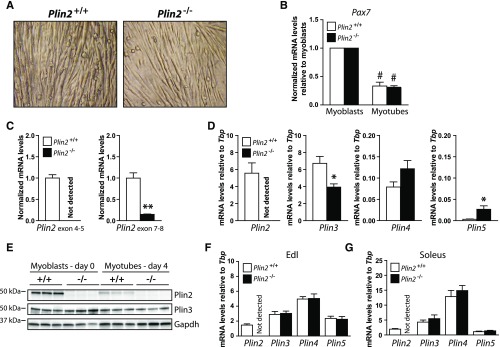
Fig. 1.
Expression of Plins in muscle and established Plin2+/+ and Plin2−/− myotubes and myoblasts. Primary muscle satellite cells (myoblasts) were isolated from the hind leg of Plin2+/+ and Plin2−/− mice. A: Established Plin2+/+ and Plin2−/− myoblast cultures differentiated equally well into multinucleated myotubes. B: Expression of Pax7 mRNA in relation to the expression of TATA-box binding protein (Tbp) determined by RT-qPCR. The results are presented normalized to the expression levels in undifferentiated myoblasts. C: RT-qPCR with primers amplifying across the Plin2 exon 4–5 junction and the Plin2 exon 7–8 junction in relation to the expression of Tbp and normalized to the expression levels in Plin2+/+ myotubes, confirmed the absence of exon 4-6 Plin2 mRNA sequences in Plin2−/− myotubes. D: Expression of Plin2, Plin3, Plin4, and Plin5 mRNAs determined by RT-qPCR in relation to the expression of Tbp. Results in B–D are presented as means ± SEM (n = 3–6, *P < 0.05 and **P < 0.01 vs. Plin2+/+ myotubes, #P < 0.05 vs. myoblasts). E: Expression of Plin2 and Plin3 proteins in myoblasts (day 0) and differentiated myotubes (day 4). The membrane contains samples from three independent experiments (n = 3). F: Relative mRNA expression of Plin2, Plin3, Plin4, and Plin5 in extensor digitorum longus of chow-fed 12-week-old Plin2+/+ and Plin2−/− male mice. G: Relative mRNA expression of Plin2, Plin3, Plin4, and Plin5 in soleus. Gene expression levels in F and G were determined by RT-qPCR and are presented in relation to the expression of Tbp as means ± SEM (n = 9 in each group). Edl, extensor digitorum longus; Pax7, paired box 7.

