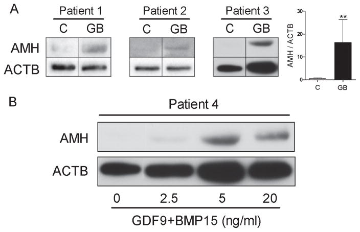
Figure 2. AMH protein levels in human cumulus cells treated with GDF9 and BMP15.
A) Cumulus cells from three different patients (Patient 1, 2, and 3) were treated with 5 ng/ml of both GDF9 and BMP15 (GB) for 48 hours. AMH β-actin (ACTB) protein levels were determined by Western blotting. On the left, the average (± SEM) of the relative optical density units of AMH to ACTB is shown (**P< 0.001, t-test, n=3). B) Cumulus cells from Patient 4 were treated with 2.5, 5, or 20 ng/ml of both GDF9 and BMP15. AMH protein levels were determined as in A. Expression of ACTB is shown as a loading control.

