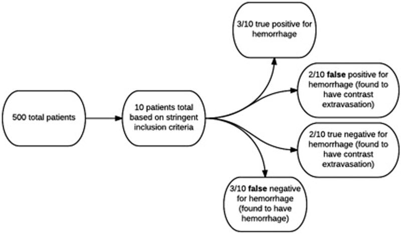
Fig 2.
In total, 500 eligible patients were identified with a database search in this retrospective study. Of those 500 patients, 10 patients qualified based on the stringent imaging inclusion criteria (Fig 1). Three out of 10 patients had a working diagnosis of postprocedural hemorrhage and were deemed true positives. Two out of 10 patients had a working diagnosis of postprocedural hemorrhage but were found to have contrast extravasation and were characterized as false positives. Two of 10 patients had a working diagnosis of contrast extravasation and were stratified as true negatives. Finally, three out of 10 patients had a working diagnosis of contrast extravasation but were found to have hemorrhage. These cases were deemed false negatives.

