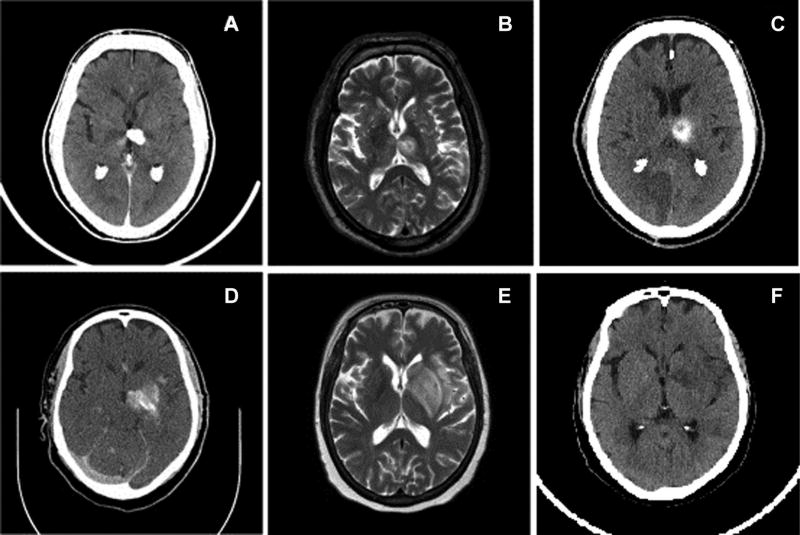
Fig 3.
Representative images of two patients (patient 1 on top row, patient 2 on bottom row) with hyperdense lesions on CT postrecanalization procedure. CT image of patient 1, a 64-year-old male, with hyperdense lesion in the left thalamus with working diagnosis of contrast extravasation with hemorrhage not excluded (A). Axial T2-weighted MRI of patient 1 demonstrating a heterogeneously hyperintense lesion centered in the left thalamus (B). Seventy-two hour postprocedural image demonstrates persistent hemorrhage in the left thalamus (C). Patient 1 was deemed to have intracranial hemorrhage. CT image of patient 2, a 57-year-old female, with hyperdense lesion in left basal ganglia and insula with working diagnosis of hemorrhage with contrast extravasation not excluded (D). Axial T2-weighted MRI of patient 2 demonstrating a heterogeneously hyperintense lesion centered in the left basal ganglia, caudate nucleus, and insula (E). Seventy-two hour postprocedural CT image demonstrating complete resolution of the previously visualized hyperdensity within the left basal ganglia (F). Patient 2 was deemed to have contrast extravasation.

