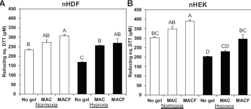
Figure 3.

The effect of oxygenating MACF hydrogels on cell metabolism, as determined by the PrestoBlue assay at day 3. A. Neonatal human dermal fibroblasts (nHDFs) and B. neonatal human epidermal keratinocytes (nHEK) cultured for 3 d with treatments. MACF improved cell metabolism under both hypoxic (1% O2) and normoxic (21% O2) environments, as evidenced by increased reducing power per sample (equivalent DTT concentration). Letters are significantly different from one another by single-factor ANOVA (p < 0.0001). Data are reported as mean ± SD with n = 3–4.
