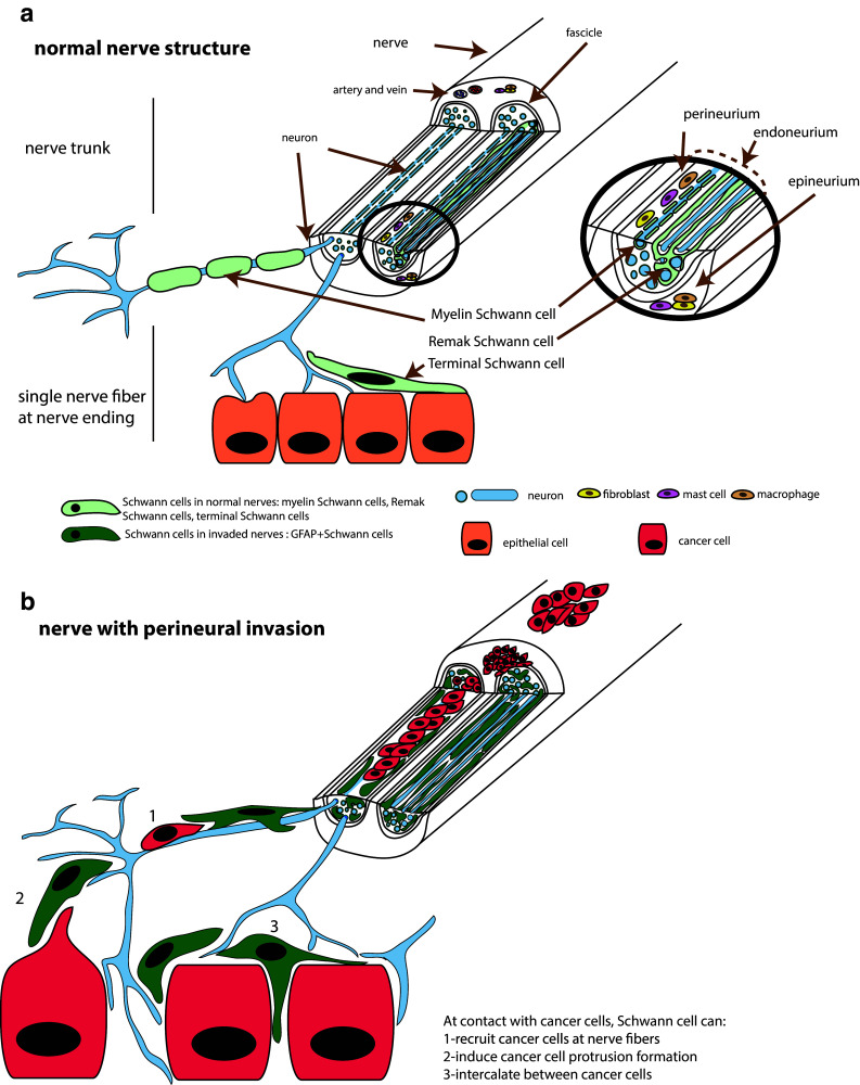
Fig. 1 .

Schematic representation of nerve structure and Schwann cells distribution. (a) Normal nerve structure showing a large caliber nerve trunk and single nerve fibers at contact with epithelial cells. Myelin, Remak and terminal Schwann cells are shown (b) Nerve with perineural invasion. Cancer cells are present around or in the nerve. At contact with cancer cells, Schwann cells can recruit cancer cells at nerve fibers, induce cancer cell protrusion formation and intercalate between cancer cells
