
Fig. 1.
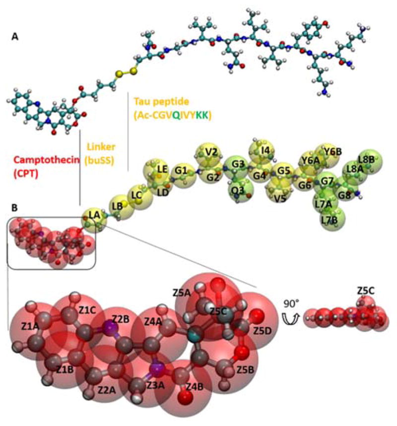
Coarse-grained and all-atomistic representations of the drug-amphiphile (DA) filaments. A. Atomistic model for ‘mCPT-buSS-Tau’, the DA in a CPK representation. B. CGed model for the DA shown as transparent VDW spheres, overlapping the atomistic model. The hydrophobic cancer drug camptothecin (CPT) is in red, the charged lysine and polar glutamine groups are shown in light green, while the rest residues are shown in yellow. CPT is expanded and rotated 90° to show the planar structure.