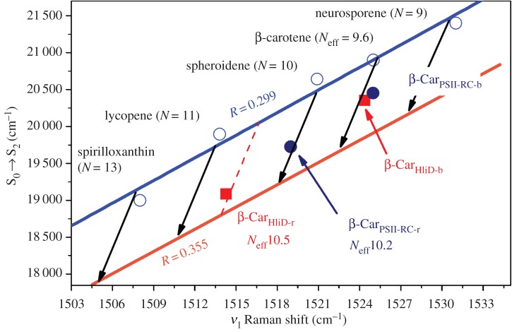
Figure 9.
Correlation between the position of the S0 → S2 electronic transition and the ν1 Raman band for β-carotenes in PSII-RC (dark blue circles) and HliD proteins (red squares) at room temperature. For comparison, the relationship between carotenoids of different conjugation lengths in the same solvent (n-hexane) is added as well as the relationship as a function of solvent polarizability (black arrows). The β-CarPSII-RC-b point corresponds to the blue-absorbing β-carotene in PSII-RC, the β-CarPSII-RC-r point corresponds to the red-absorbing β-carotene in PSII-RC, the β-CarHliD-b point corresponds to the blue-absorbing β-carotene in HliD, and the β-CarHliD-r point corresponds to the red-absorbing β-carotene in HliD.

