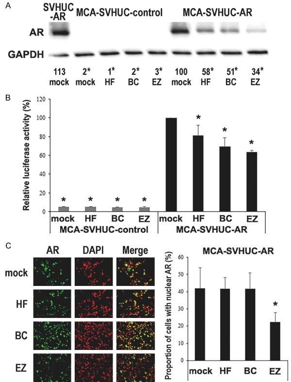
Figure 1.

Effects of enzalutamide on AR in MCA-exposed urothelial cells. A. Cell extracts from SVHUC-AR without MCA exposure or SVHUC-control/SVHUC-AR exposed to MCA and subsequently treated with ethanol (mock), hydroxyflutamide (HF; 5 μM), bicalutamide (BC; 5 μM), or enzalutamide (EZ; 5 μM) for 48 hours were analyzed on western blotting, using an antibody to AR (110 kDa). GAPDH (37 kDa) served as an internal control. The means of densitometry values for AR standardized by GAPDH that are relative to the value of mock treatment (6th lane; set as 100%) from three independent experiments are included below the lanes. B. Luciferase reporter gene assay was performed in SVHUC-control or SVHUC-AR exposed to MCA, transfected with MMTV-Luc and pRL-TK, and subsequently treated with ethanol (mock), hydroxyflutamide (HF; 5 μM), bicalutamide (BC; 5 μM), or enzalutamide (EZ; 5 μM) for 24 hours. Luciferase activity is presented relative to that of mock treatment in SVHUC-AR cells. Each value represents the mean + standard deviation from three independent experiments. C. Immunofluorescence of AR in SVHUC-AR cells exposed to MCA and subsequently treated with ethanol (mock), hydroxyflutamide (HF; 5 μM), bicalutamide (BC; 5 μM), or enzalutamide (EZ; 5 μM) for 24 hours. We merged the images between AR and DAPI that was used to visualize nuclei, and the proportion of cells with AR expression only in their nuclei was quantitated. Each value represents the mean + standard deviation of 6 determinants. *P < 0.05 (vs. mock treatment in SVHUC-AR).
