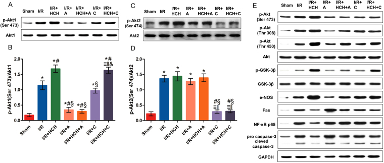
Figure 5.
The roles of Akt1 and Akt2 in the protective effects of HCH in experiment 1. Mice were sacrificed after 4 h of reperfusion and the hearts were harvested for biochemical examinations (n = 56). The protein expression of p-Akt1 (Ser 473), Akt1, p-Akt2 (Ser 474), Akt2, p-Akt (Ser 473), p-Akt (Thr 308), p-Akt (Thr 450), Akt, p-GSK-3β, GSK-3β, e-NOS, Fas, NF-κB p65, pro-caspase-3, and cleaved caspase-3 was detected by western blotting (A,C,E). p-Akt1 expression increased markedly in the heart after 90-min HCH treatment. However, it was reduced significantly in the I/R + HCH + A group (B). There was no significant difference in p-Akt2 (Ser 474) among the I/R, I/R + HCH, I/R + HCH + A, and I/R + A groups, but p-Akt2 (Ser 474) levels were significantly decreased in the I/R + HCH + C and I/R + C groups (D). *P < 0.05 vs. sham; # P < 0.05 vs. I/R; § P < 0.05 vs. I/R + HCH; ≡ P < 0.05 vs. sham; ‖ P < 0.05 vs. I/R; & P < 0.05 vs. I/R + HCH.

