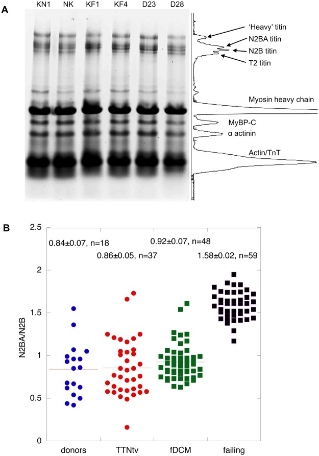
Figure 3.
Titin isoform expression. (A) 2% agarose/2% polyacrylamide-SDS gel electrophoresis of human heart myofibrils showing separation of titin isoforms, identified in the scanned profile: Two donors, KN1, NK, two failing heart, KF1, KF4, and two TTNtv mutant samples are shown. (B) Comparison of N2BA/N2B ratio measured in donors, TTNtv, other fDCM and idiopathic DCM. Pooled data from multiple gels similar to A. Data shown for donor heart represents KN1 (17 replicates), NK (13 replicates) and NH (8 replicates). Data shown for TTNtv represents D6 (7 replicates), D7 (8 replicates), D9 (6 replicates), D23 (15 replicates), D28 (15 replicates) and D29 (4 replicates). Data for other fDCM represents D1 (5 replicates), D2 (8 replicates), D4 (4 replicates), D11 (9 replicates), D12 (9 replicates) and D15 (9 replicates). Data shown for idiopathic failing heart represents FG (13 replicates), FH (11 replicates), FI (7 replicates), KF1 (8 replicates), KF2 (3 replicates), KF3 (9 replicates) and KF4 (12 replicates). Only idiopathic DCM samples have a significantly higher N2BA/N2B ratio than donor (1.9x).

