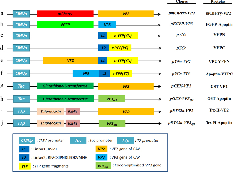
Figure 1.
Schematic diagram of the constructs used for expression of VP2 and Apoptin of CAV. Constructs a and b used for investigation of the subcellular distribution of VP2 and Apoptin are indicated as the VP2 gene and VP3 gene, which were fused with fluorescent mCherry and EGFP, respectively, in the transient expression vectors pmCherry-C1 and pEGFP-C2 to generate the recombinant plasmids pmCherry-VP2 and pEGFP-VP3, respectively. Constructs c and d were used as negative controls in ex vivo BiFC assays and indicate that the two gene fragments of YFP encoding the N-terminal domain (YN, residues 1–154) and C-terminal domain (YC, residues 155–238) were respectively cloned into plasmid pcDNA3.1 for cell transfection. Linker 1 (L1) and linker 2 (L2) represent sequences encoding two small peptides (RSIAT for L1 and RPACKIPNDLKQKVMNH for L2) used for the linkage of YN and YC. The VP2 and VP3 genes were respectively fused to the sequences of YN and YC using L1 and L2 as linkers, resulting in constructs e and f. Constructs g, h, i and j were used for in vitro GST pull-down assays and were generated for expression of VP2 and Apoptin of CAV by fusion of the VP2 and VP3 genes. Constructs g and h were generated from pGEX-4T-1 for GST fusion protein expression. The thioredoxin-His (Trx-H) tag for VP2 and apoptin was obtained via expression of the Trx-H coding sequence from plasmid pET32a, illustrated as constructs i and j, respectively.

