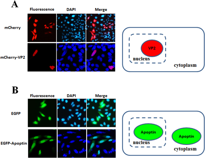
Figure 2.
Subcellular localisation of CAV VP2 and Apoptin in CHO-K1 cells. CHO-K1 cells were transfected with pmCherry-C1 and pmCherry-VP2 (A) or pEGFP-C2 and pEGFP-VP3 (B), respectively. At 48 hrs post-transfection, the cells were fixed and stained with DAPI as a nuclear indicator, followed by confocal microscopy. The distribution of red fluorescence in CHO-K1 cells indicated the subcellular localisation of overexpressed mCherry or mCherry-VP2; green fluorescent indicated the localisation of EGFP or EGFP-Apoptin. The localisation of VP2 and Apoptin in CHO-K1 cells was determined after comparing the merged images between the sole fluorescent protein and fluorescent protein-fused protein.

