Fig. 4.
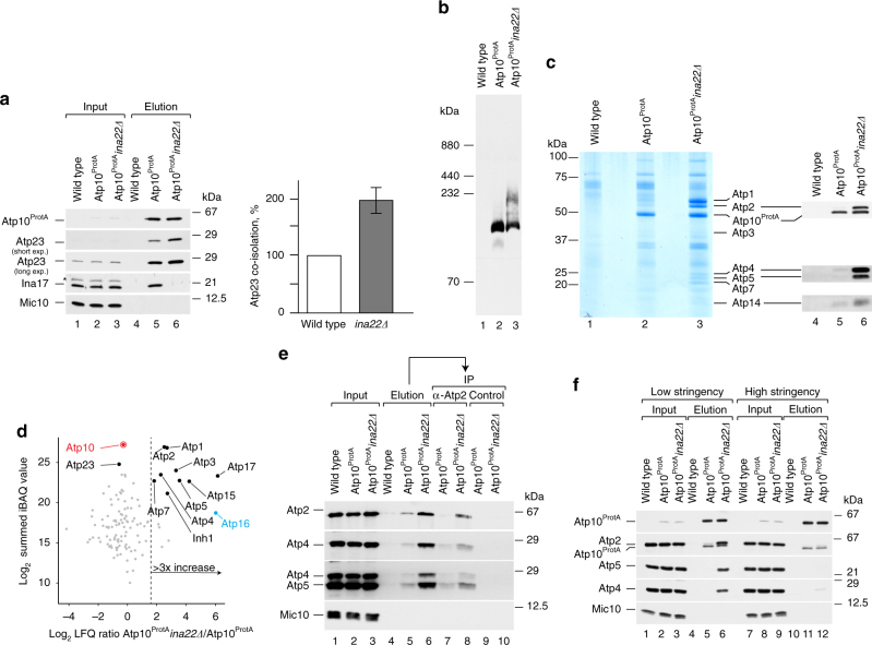
Atp10 accumulates with Atp23, peripheral stalk and F1 module in ina22Δ. a Atp10ProtA-containing protein complexes were purified via IgG-affinity chromatography from Atp10ProtA, Atp10ProtA ina22Δ, and wild-type mitochondria. Input and elution fractions were analyzed by SDS-PAGE and western blotting with indicated antibodies (left panel). The amount of Atp23 isolated from Atp10ProtA mitochondria was set as 100% and Atp23 co-isolated from Atp10ProtA ina22Δ mitochondria was quantified (right panel). Signals were normalized to the amounts of isolated Atp10ProtA. Input = 1% of elution. (n = 3, ±SEM). The asterisk (*) indicates a cross-reacting band. b The experiment was performed as in a, bound complexes were eluted natively and analyzed by BN-PAGE and western blotting with anti-Atp23 antibodies. c Wild type, Atp10ProtA, and Atp10ProtA ina22Δ mitochondria were subjected to IgG-affinity chromatography. Ninety percent of the elution fractions was analyzed by SDS-PAGE and colloidal Coomassie staining (lanes 1–3). Remaining fractions were analyzed by SDS-PAGE and western blotting with indicated antibodies (lanes 4–6). d Atp10ProtA-containing protein complexes were purified via IgG-affinity chromatography from Atp10ProtA and Atp10ProtA ina22Δ mitochondria. Eluates were analyzed by BN-PAGE followed by LC-MS and label-free quantification using the MaxQuant LFQ and the iBAQ algorithm. The scatter plot shows for each protein the logarithmic (log2) ratio of LFQ intensities for Atp10ProtA ina22Δ over Atp10ProtA plotted against the summed iBAQ intensity of Atp10ProtA ina22Δ and Atp10ProtA. The dashed line indicates a non-logarithmic ratio of three. Red, bait protein; blue, Atp16 was only identified in Atp10ProtA ina22Δ and the ratio was set to six. e Wild type, Atp10ProtA, and Atp10ProtA ina22Δ mitochondria were subjected to IgG-affinity chromatography. Protein complexes were eluted natively via TEV-cleavage. Elution fractions were subjected to immunoprecipitation (IP) with either anti-Atp2 antibodies or a control pre-immune serum. 1% of the input fraction and entire elution fraction were analyzed by SDS-PAGE and western blotting with indicated antibodies. f Wild type, Atp10ProtA, and Atp10ProtA ina22Δ mitochondria were solubilized in a high- or low-stringency buffer (see “Experimental procedures”) and subjected to IgG-affinity chromatography. Input and elution fractions were analyzed by SDS-PAGE and western blotting with indicated antibodies. Input = 1% of elution

