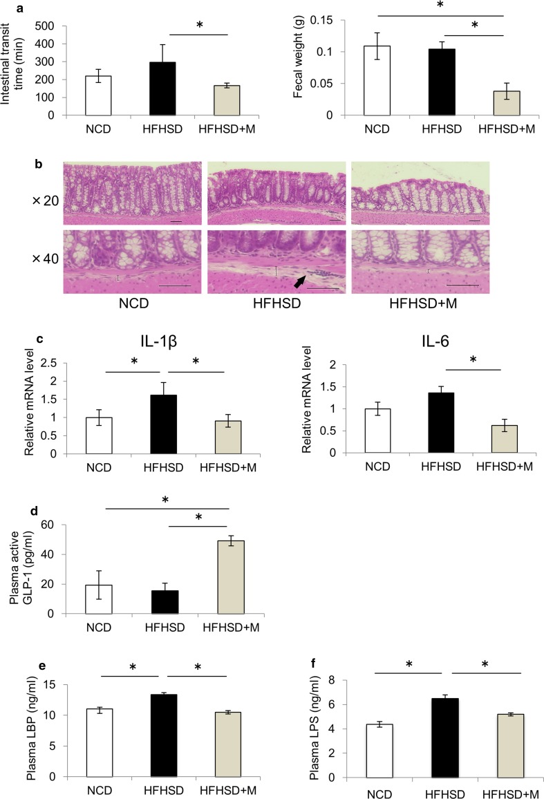
Fig. 3.
Miglitol changed the gut environment and reduced endotoxemia. Mice were divided into three groups and fed a normal chow diet (NCD), a high-fat high-sucrose diet (HFHSD), or HFHSD containing 0.04% miglitol (HFHSD+M) for 12 weeks and then killed. a The intestinal transit times were determined by measurement of the time elapsed and the color of excreted feces. The colonic fecal weight was determined at the time when the mice were killed (n = 10 per group). b Colon sections were stained with hematoxylin–eosin. Scale bar 50 μm (×20, ×40 magnification). The straight line indicates the extent of the submucosal edema and arrows indicate inflammatory infiltrates composed primarily of neutrophils. c Colonic messenger RNA (mRNA) levels of IL-1β and IL-6 (n = 4–5 per group). d Plasma active glucagon-like peptide 1 (GLP-1) levels (n = 6–8 per group). e Plasma lipopolysaccharide-binding protein (LBP) levels (n = 6–8 per group). f Plasma lipopolysaccharide (LPS) levels (n = 6–7 per group). Data are presented as the mean ± standard error of the mean. Asterisk statistical significance (p < 0.05)

