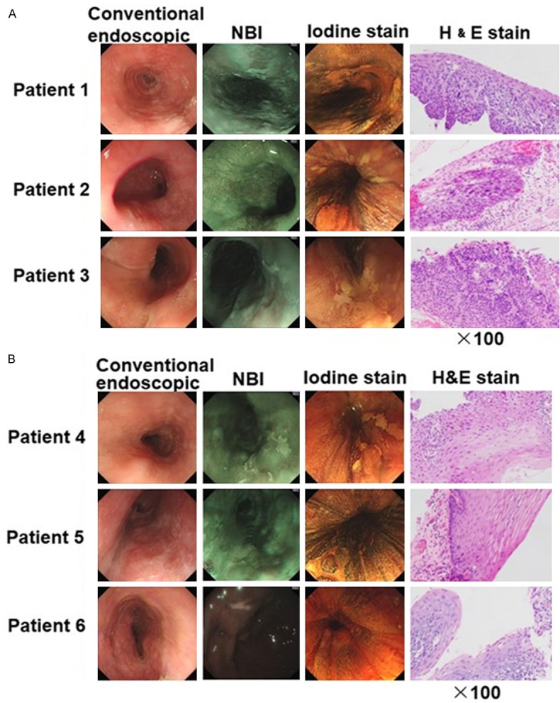
Figure 1.

Routine endoscopy and narrow band imaging (NBI) images of representative cases of HGIN patients and LGIN patients. Conventional endoscopic view: a slightly depressed lesion with iodine unstained areas. Resected specimens with H&E (×100) stain were shown. A. Specimens were diagnosed as HGIN if stained with neoplastic cells > 50% in the thickness of the epithelium. B. Specimens were diagnosed as LGIN if stained with neoplastic cells < 50% in the thickness of the epithelium.
