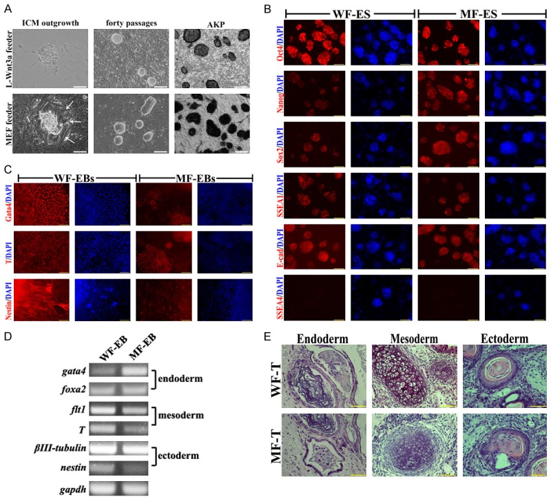
Figure 1.

Pluripotent analysis of ES cells on MEFs and L-Wnt3a cells feeder layer. A. Blastocyst outgrowth on L-Wnt 3a cell and MEFs feeder layers, morphology of WF-ES and MF-ES cells, and AKP staining, bar=100 μm; B. Immunostaining of Oct4, Nanog, Sox2, SSEA1, SSEA4 and E-cadherin in WF-ES and MF-ES cells, bar=100 μm. C. Immunostaining of Gata4, T and Nestin in EBs that derived from WF-ES and MF-ES cells, bar=100 μm; D. Expression of three germ layer genes in EBs that derived from WF-ES and MF-ES cells; E. Tertomas from WF-ES and MF-ES cells, bar=50 μm.
