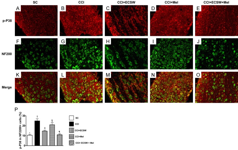
Figure 7.

Immunofluorescent (IF) microscopic finding of colocalization of p-P38 and NF200 indorsal root ganglion (DRG) neurons. A-E. Illustrating the IF microscopic finding (200x) positively stained p-P38 in DRG neurons (red color spots). F-J. Illustrating the IF microscopic finding (200x) positively stained NF200 cells (green color). K-O. Illustrating the IF microscopic finding (200x) of merged positively-stained p-P38 and NF200 (green-red colocalization). P. Analytical results of number of p-P38+/NF200+ cells, * denotes statistical significance vs. other groups with different symbols (†, ‡, §, ¶), p<0.0001. All statistical analyses were performed by one-way ANOVA, followed by Bonferroni multiple comparison post hoc test (n = 8 for each group). Symbols (*, †, ‡, §, ¶) indicate significance at the 0.05 level. SC = sham control; CCI = chronic constriction injury; ECSW = extracorporeal shock wave; Mel = melatonin.
