
Figure 5.
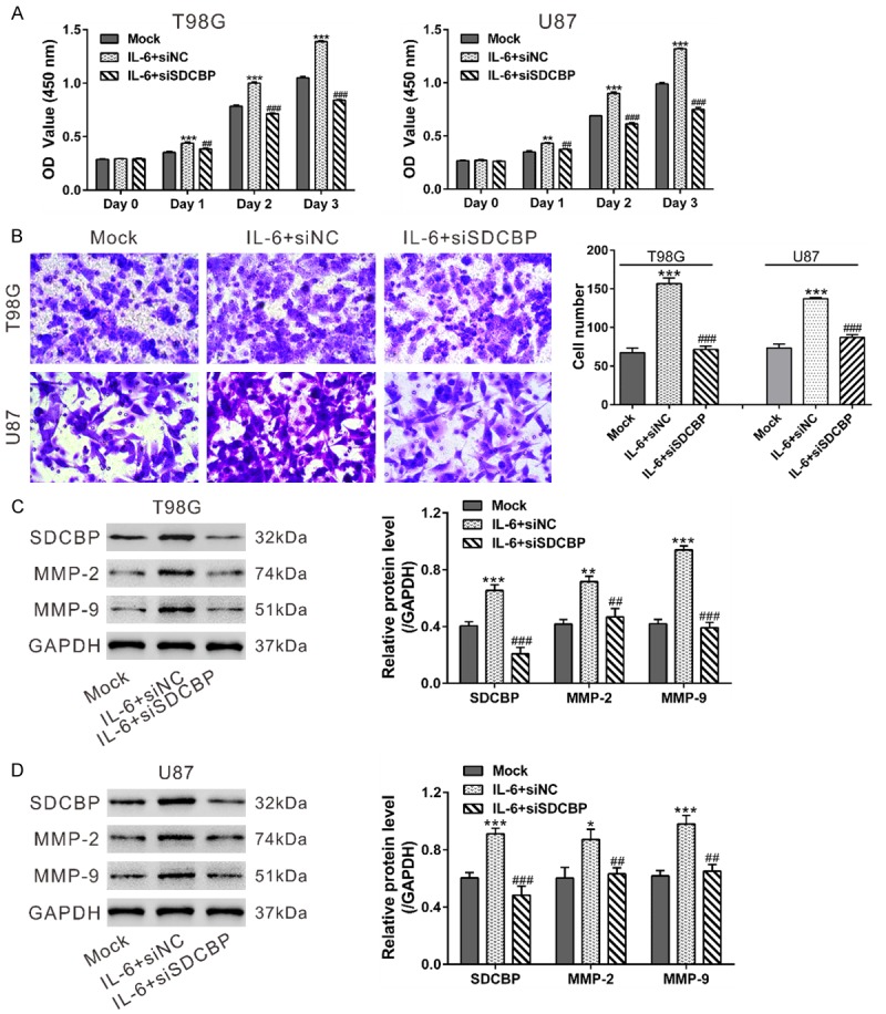
IL-6 promoted glioma cell growth and invasion via SDCBP. T98G and U87 cells were transfected with siSDCBP or siNC. At 24 h after transfection, cells were serum-starved overnight. A. Cells were seeded onto 96-well plates for CCK-8 assays to measure cell proliferation in the presence of IL-6 (100 ng/mL). Untreated cells (Mock) was used as a control. B. Cells were seeded onto the upper chamber containing MEM, and the lower chamber was filled with MEM+100 ng/mL IL-6. Transwell assays were used to assess the invasive potential of glioma cells after 24 h. C, D. Expression of SDCBP, MMP-2, and MMP-9 were determined in cells treated in the same manner as in the Transwell assays. *P<0.05, **P<0.01, and ***P<0.001 vs. Mock; ##P<0.01 and ###P<0.001 vs. IL-6+siNC.