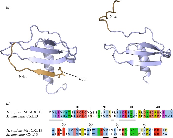
Figure 2.
Comparison of human Met-CXCL13 and murine wt-CXCL13 sequences and structures. (a) Structure of human Met-CXCL13 (left, PDB ID 4ZAI) and murine wt-CXCL13, in which the initiating Met residue was removed (right, PDB ID 5L7M), depicted as cartoon, illustrating the differences in N-terminal domain (in brown) folding. (b) Sequence alignment of the human and murine CXCL13, used for structure resolution, depicted with a colour and intensity code according to residue type and homology (prepared with ClustalW). The residues involved in the N-terminal domain folding (1–4, 23, 26–31, 65–66 and 69) are underlined.

