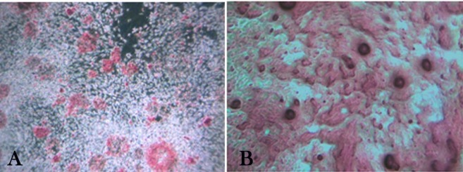
Figure 3.
Osteogenic differentiation of deciduous and permanent periodontal ligament stem cells (stained with alizarin red on day 21 of culture). Bone nodule-like structures under an inverted light microscope, indicating calcium deposition in both groups of cell cultures; a) Deciduous periodontal ligament stem cell (×100); b) Permanent periodontal ligament stem cells (×200 magnification).

