Figure 2.
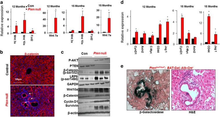
Wnt signaling is induced in steatotic Pten-null livers. (a) Quantitative PCR analysis for the expression of Wnt ligands and receptors in livers of Pten-null and control mice. n=5. * Indicates significant difference from controls at P⩽0.05. (b) β-catenin staining (red) in control and Pten-null mice. β-catenin is expressed throughout the normal liver. Its levels are increased in the Pten-null livers at the hyperplastic ductal region where the progenitor cell niche is located (arrow). Blue, 4',6-diamidino-2-phenylindole (DAPI). Representative of three animals. (c) Immunoblotting analysis of PTEN signal and Wnt signal in livers from 12-month-old mice. Glyceraldehyde 3-phosphate dehydrogenase (GAPDH) and β-actin are loading controls. (d) Quantitative PCR analysis of Wnt target genes in control and Pten-null livers. n=3–5. * Indicates significant difference from controls at P⩽0.05. (e) β-galactosidase activity was detected in the periductal area (dark greenish blue staining in left panel) in the 6-month-old Pten-null liver carrying BAT-Gal reporter construct. Right panel, hemotoxylin & eosin (H&E) staining in adjacent section. Representative of three animals.

