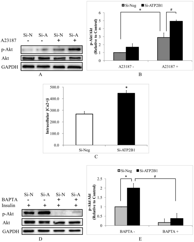
Figure 2.
The increased insulin sensitivity in ATP2B1-silenced HUVECs cells may be due to calcium-dependent signaling pathway. HUVECs were starved of serum overnight before stimulation with 500 nM A23187 for 30 min. A. Representative Western blotting results of p-Akt, Akt and GAPDH(n=4). B. Fold changes of phosphor-Akt vs. total Akt relative to its basal levels in the control cells without A23187 stimulation were quantified by densitometry. *P<0.05, vs. negative siRNA-transfected cells without A23187 stimulation. #P<0.05, vs. negative siRNA-transfected cells stimulated with A23187. C. The intracellular concentrations of Ca2+ were measured using fluo-3/AM in HUVECs 48 h after transfection(n=6). *P<0.05, vs. cells transfected with negative siRNA. Following 2 hours of BAPTA administration HUVECs were stimulated with 100 nM insulin for 30 min. D. Representative Western blotting results of phosphor-Akt (Ser473) (n=4). E. Fold changes of phosphor-Akt vs. total Akt relative to its basal levels in control cells were quantified by densitometry. *P<0.05, vs. cells transfected with negative siRNA; #P<0.05, vs. ATP2B1-silenced cells without BAPTA-AM pretreatment.

