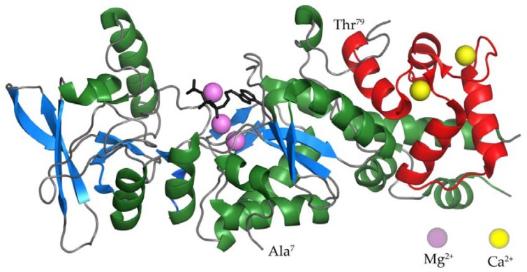
Figure 2.
The crystal structure of the AC domain of CyaA in the complex with the C-terminal domain of CaM [53]. α-helices and β-strands of the AC domain are colored in green and blue, respectively. The C-terminal domain of CaM is colored in red. Calcium and magnesium ions are represented by yellow and violet balls. The structure of the ATP analog adefovir diphosphate is represented by black lines.

