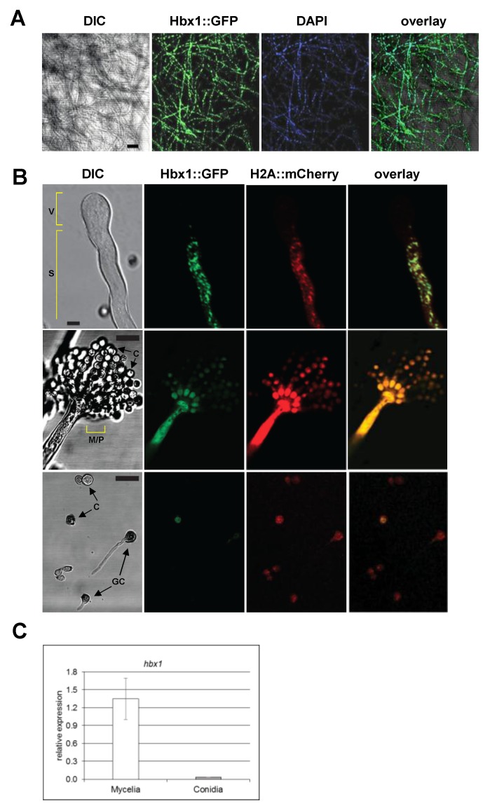
Figure 7.
Localization of Hbx1 protein using CLSM. (A) Analysis of spatial distribution of Hbx1 within mycelia. CA14 expressing Hbx1::GFP was grown on solid PDA growth medium and imaged at 48 h time point. Scale bar, 50 μm. (B) Localization of Hbx1 in developing conidiophores. Upper panels: imaging of a conidiophore initiating vesicle formation. CA14 expressing Hbx1::GFP and histone H2A::mCherry was grown on PDA and studied after 48 h. Note lack of Hbx1 and nuclei in the vesicular region. Middle panels: imaging of a mature conidiophore. Note decrease in Hbx1 localization to conidia. Scale Bar, 5 μm; Bottom panels: Conidia (103) of A. flavus CA14 expressing Hbx1::GFP and histone H2A::mCherry were inoculated in 10 µL of potato dextrose broth and were observed under CLSM 6 h after inoculation (a time that corresponds to the initiation of spore germination). For both samples, the image acquisition conditions were adjusted such that Hbx-1:GFP could be visualized in at least one conidium. The adjusted image conditions were then propagated to red channel (to visualize H2A::mCherry) as well. Fluorescence intensity was increased in the middle and lower panels in order to clearly see fluorescence in the metulae/phialides and conidiospores. Scale bar, 10 µm. (C) RT-qPCR analysis of hbx1 expression levels in CA14 control grown with agitation in WKMU broth for 2 d (mycelia) or WKMU agar plates for 6 d (isolated conidia). Abbreviations: GC, germinating conidium; C, conidium; M/P, metular/phialide region; V, vesicle; S, stipe.

