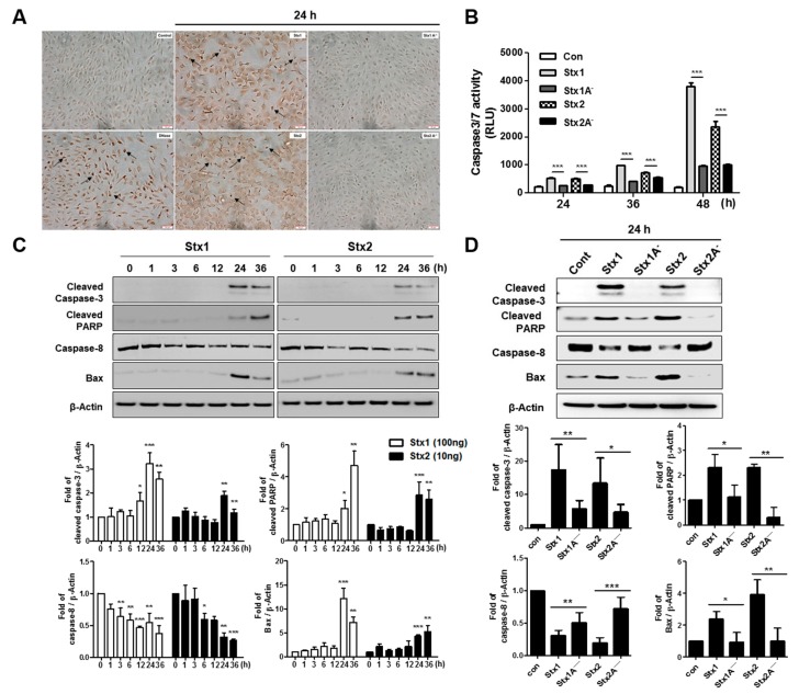
Figure 6.
Stxs activate apoptotic signaling pathways in RPE cells. (A) The TUNEL assay was performed to detect apoptotic APRE-19 cells after treating with Stx1, Stx2, Stx1A− or Stx2A− for the indicated times. Cells staining dark brown are TUNEL positive and are indicated by arrows. Scale bars = 50 μm; (B) Analysis of caspase-3/7 activity. ARPE-19 cells were incubated with Stx1 (100 ng/mL), Stx1A− (100 ng/mL), Stx2 (10 ng/mL) and Stx2A− (10 ng/mL) for the indicated time points, and caspase 3/7 activity was measured using the Caspase-Glo 3/7 Assay; (C,D) ARPE-19 cells (1.0 × 10⁵ cells/well) were treated with Stx1 (100 ng/mL), Stx1A− (100 ng/mL), Stx2 (10 ng/mL) and Stx2A− (10 ng/mL) for the indicated time points. Protein samples were prepared and analyzed by Western blotting using anti-cleaved caspase-3, anti-caspase-8, anti-cleaved PARP, anti-Bax and anti-β-Actin antibodies. β-Actin was used as a control for equal protein loading. The results are from one representative experiment of three independent experiments. The bar graphs show the mean ± SEM of fold changes in band densities normalized by division of β-Actin band densities and compared to untreated control cell values (lower panels). Asterisks indicate significant differences between control cell values vs. Stxs-treated cells (panel C) or Stx 1 vs. Stx1A− and Stx2 vs. Stx2A− (panel D); * = p < 0.05; ** = p < 0.01; *** = p < 0.001.

