
Figure 3.
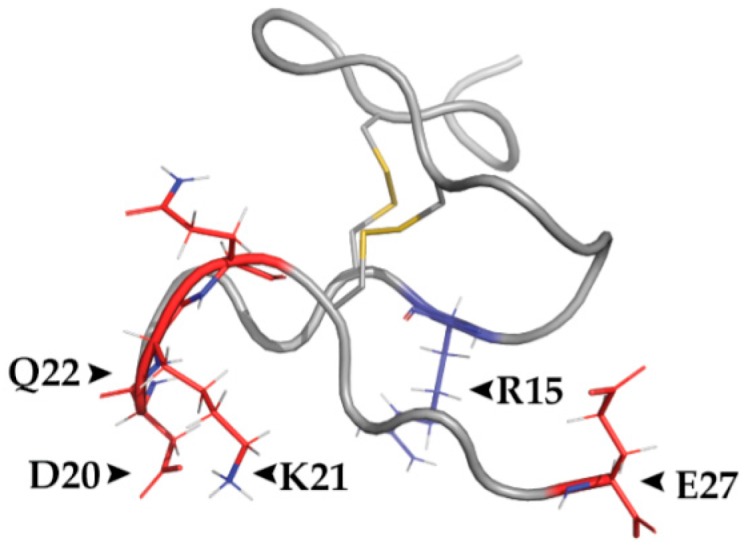
The polar RhTx binds TRPV1 through its charged surface. Structure of RhTx indicating the polarity of the molecule. Charged residues D20, K21, Q22 and E27 (red) have been indicated in TRPV1 binding, along with the polar residue R15. Two cysteine bridges are highlighted in yellow. PDB ID: 2MVA.