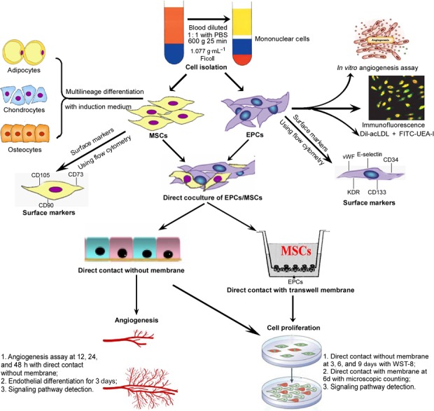
Figure 7.

A schematic flow diagram of this study. hEPCs and hMSCs were isolated from human bone marrow. The isolated hEPCs were characterized with in vitro angiogenic potential and the specific markers. The isolated hMSCs were characterized with the multilineage differential potential and the specific surface markers. Then hEPCs and hMSCs were cocultured in a two‐dimensional (2D) monolayer mixed or 3D transwell membrane cell‐to‐cell coculture systems. The proliferation and angiogenic capacities of cells were assessed, and the underlying mechanism was investigated.
