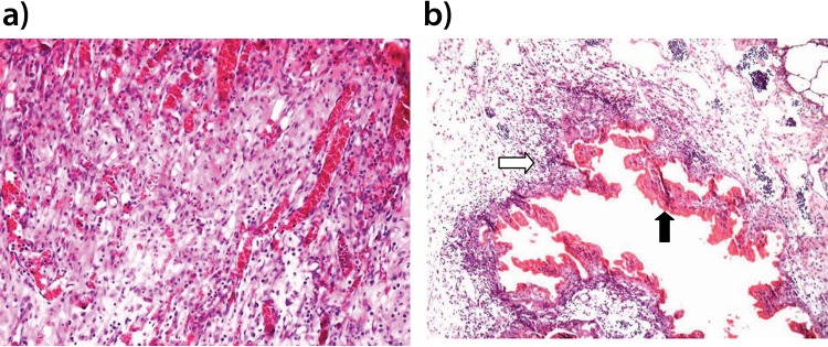
Figure 1.
Photomicroscopy image of rabbit lung parenchyma stained by HE (X100 magnification). The image in a (FB group) shows intense neoangiogenesis and inflammatory cell infiltration, which are findings compatible with normal wound healing. The image in b (CA group) shows a palisade of histiocytic cells and lymphocytes (white arrow) and a central area of necrosis (black arrow).

