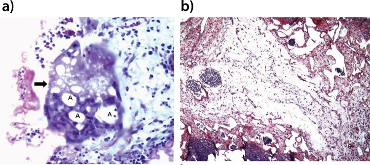
Figure 2.
Photomicroscopy image of rabbit lung parenchyma from the CA group. The image in a shows a multinucleated giant cell (arrow) with cytoplasmic vacuoles containing CA residues with HE staining (X400 magnification) (A). The image in b shows an absence of collagen deposits and accentuated stromal edema with picrosirius red staining (X100 magnification).

