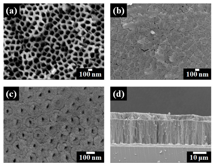
Figure 2.
Field emission scanning electron microscope (FE-SEM) images of freestanding TiO2 nanotube arrays. (a) Top view; (b) Bottom view before ion milling process; (c) Bottom view after ion milling process of freestanding TiO2 nanotube arrays; and (d) Side view of freestanding TiO2 nanotube arrays and large TiO2 NPs on the fluorine-doped tin oxide (FTO) glass.

