
Figure 1.
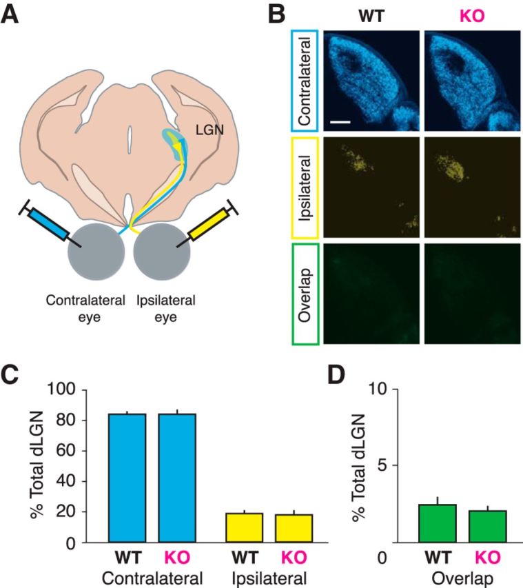
Segregation of ocular inputs to LGN is normal in juvenile Cx3cr1 KO animals. A, Schematic of eye injection procedure, where anterograde tracers (CTB Alexa Fluor conjugates) are injected into each eye, labeling RGC projections in the LGN. B, Representative images of LGN with contralateral retinal projections (blue), ipsilateral projections (yellow), and the overlapping signal of both eyes (green). C, Average of labeled LGN areas shows no difference in any measurement between genotypes (contralateral, p = 0.585; ipsilateral, p = 0.528; overlap, p = 0.482). D, Overlap between contralateral and ipsilateral projections is minimal (n = 6 WT, 10 KO animals).