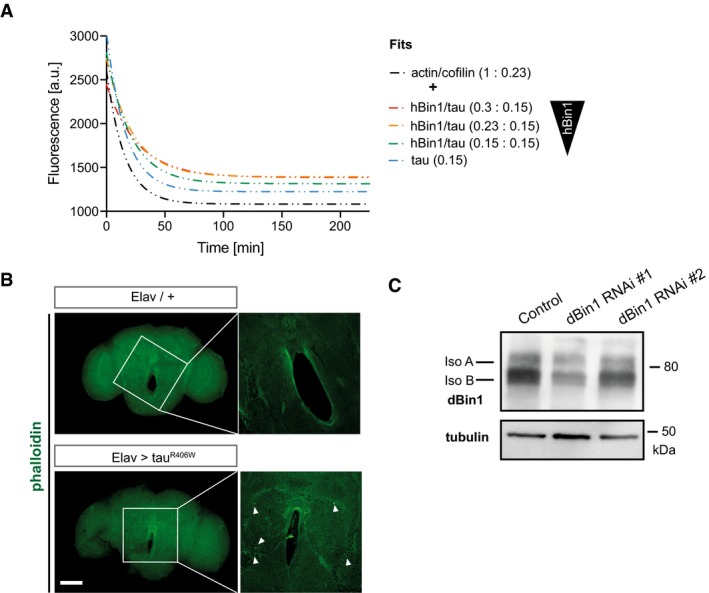
Figure EV5. Biochemical validation of dBin1 downregulation in fly brains.

- Phalloidin staining of whole‐mount brain (22 days old) dissected from tauR406W transgenic flies and control flies. The zoom‐in images represent one single section in the middle of the central body of the fly brain, and the white arrows highlight actin rods. Scale bar of brain overview: 100 μm and zoom‐in: 25 μm.
- Knockdown efficiency of different dBin1 RNAi lines. The dBin1 lines were expressed pan‐neuronally under the Elav promoter, and fly heads were subjected to protein extraction 10 days post‐eclosion. Samples were resolved by SDS–PAGE and immunoblotted.
