-
A
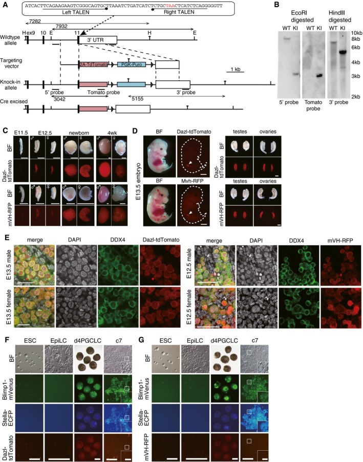
The targeting strategy to create a
Dazl‐tdTomato (DT) reporter using the TALEN system (Sakuma
et al,
2013). H: Hindlll site; E: EcoRl site.
-
B
Southern blot analysis of the Blimp1‐mVenus (BV); Stella‐ECFP (SC); and DT ESCs. The correct targeting was verified using the 5′‐, 3′‐, and tdTomato probes. WT: parental wild‐type cells; KI: BVSCDT knockin ESCs.
-
C
DT and mVH‐RFP (VR) expression in testes and ovaries at the indicated developmental stages. BF: bright field images. Scale bars, 800 μm.
-
D
(Left) Bright field and fluorescence images of DT (top) and VR (bottom) expression in whole embryos at E13.5. Note that DT and VR show specific expression in gonads (arrows). The outlines of the embryos are delineated by dotted lines. Scale bar, 2 mm. (Right) Bright field and fluorescence images of DT (top) and VR (bottom) expression in isolated testes and ovaries at E13.5. Note that DT and VR are strongly expressed in gonads but not in mesonephroi. Scale bar, 400 μm.
-
E
Co‐/specific expression of DT (left) and VR (right) in DDX4 (+) germ cells in E13.5 (left) and E12.5 (right) testes and ovaries revealed by immunofluorescence (IF) analysis. Scale bars, (left) 20 μm; (right) 50 μm.
-
F, G
Bright field and fluorescence images of Blimp1‐mVenus (BV); Stella‐ECFP (SC); DT (F) or BVSCVR (G) expression during PGCLC induction and culture. Note that DT and VR are expressed at low levels/not expressed in d4 and c7 PGCLCs (the boxed area is magnified in the inset; scale bar, 40 μm). Scale bars, 200 μm.

