Figure 5.
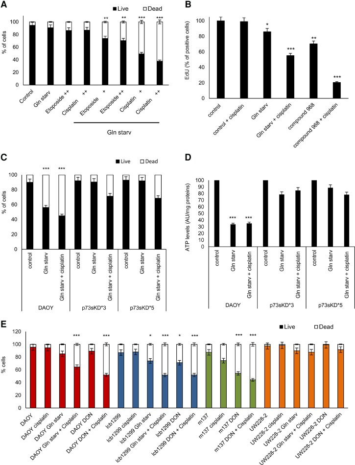
Cotreatment of cisplatin plus glutamine starvation induces a synergic apoptotic effect in MB. (A) DAOY cells were cultured for 18 h under normal medium or glutamine starvation with or without etoposide ([+] 6.8 nM; [++] 13.6 nM) or cisplatin ([+] 16.6 nM; [++] 33.3 nM). Apoptosis was measured with Annexin V-FITC and PI for flow cytometry analysis. Data are represented as mean ± SD. n = 3. (**) P < 0.001; (***) P < 0.0001. (B) Histogram shows mean fluorescence intensities of EdU in ICb-1299 cells under control medium, glutamine starvation, or 4 µM compound 968 with or without 16.6 nM cisplatin for 18 h. (*) P < 0.01; (**) P < 0.001; (***) P < 0.0001. (C,D) DAOY cells were infected with an empty vector or shRNA p73sKD*3 and p73sKD*5. Cells were incubated for 36 h under normal medium, glutamine starvation, or glutamine starvation plus 16.6 nM cisplatin. Apoptosis was measured with Annexin V-FITC and PI by flow cytometry (C) or ATP levels (D). Data are represented as mean ± SD. n = 3. (***) P < 0.0001. (E) Apoptosis was determined in DAOY, ICb-1299, m137, and UW228-2 cells. Cells were treated with 16.6 nM cisplatin, glutamine starvation, glutamine starvation plus 16.6 nM cisplatin, 0.9 mM 6-diazo-5-oxo-L-norleucine (DON), or DON plus 16.6 nM cisplatin. The cells were collected and stained with Annexin V-FITC and PI for flow cytometry analysis. Data are represented as mean ± SD. n = 3. (*) P < 0.01; (***) P < 0.0001.

