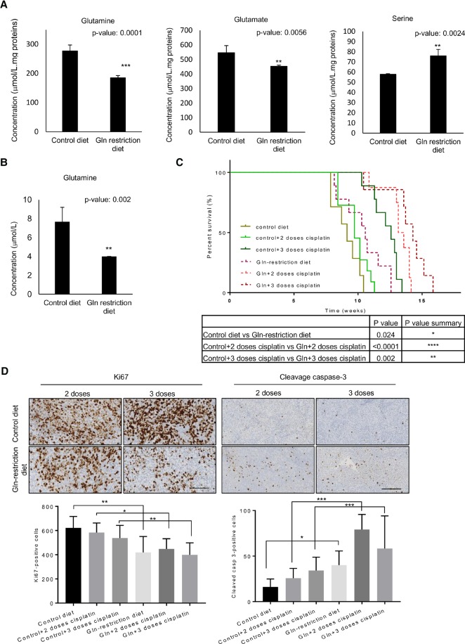
Figure 7.
A glutamine restriction diet induces a significantly improved survival in an orthotopic MB xenograft model. (A,B) NOD-SCID mice were treated for 4 mo with a control diet or glutamine restriction diet. (A) Glutamine, glutamate, and serine levels were determined by ion-exchange chromatography in the cerebellum. (**) P < 0.001; (***) P < 0.0001. (B) Glutamine level was determined by ion-exchange chromatography in the CSF. Data are represented as mean ± SD. n = 5. (**) P < 0.001. (C,D) ICb-1299 human primary cells were injected into the cerebellum of newborn NOD-SCID mice, and mice were divided into control diet (n = 7), control diet + two doses of cisplatin (n = 11), control diet + three doses of cisplatin (n = 9), glutamine restriction diet (n = 9), glutamine restriction diet + two doses of cisplatin (n = 8), or glutamine restriction diet + three doses of cisplatin (n = 7). (C) The survival of the mice is plotted over time (log-rank test). (*) P < 0.01; (**) P < 0.001; (****) P < 0.00001. (D) Histology of the MB tumors under a control diet with two or three doses of cisplatin or under a glutamine restriction diet with two or three doses of cisplatin. Representative bright-field images are shown for Ki-67 and cleaved caspase-3. Quantification is shown as the mean of positive cells per high-power field. Bar, 50 µm. (*) P < 0.01; (**) P < 0.001; (***) P < 0.0001.

